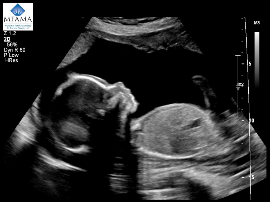
When Is an Ultrasound Performed During Pregnancy?Īn ultrasound is generally performed for all pregnant women around 20 weeks into their pregnancy. Ultrasound does not use radiation, as other procedures, such as X-rays, do. Done properly means it's performed by a physician or a trained technician, called a sonographer. But, there's no evidence to show a prenatal ultrasound done properly will harm a mother or their unborn child.

Is Prenatal Ultrasound Safe?Īll medical procedures have risk. Transvaginal ultrasound is also used to evaluate the cervix for problems such as shortening which may increase your risk of early labor. It can also be used to determine how far along you are in your pregnancy (gestational age). It may be used early in pregnancy to get a clearer view of the uterus or ovaries if a problem is suspected. This method of ultrasound produces an image quality that is greatly enhanced. However, a transvaginal ultrasound is an alternative procedure in which a tubular probe is inserted into the vaginal canal. Most prenatal ultrasound procedures are performed topically, or on the surface of the skin, using a gel as a conductive medium to aid in the image quality.

Major anatomical abnormalities or birth defects may be visible on an ultrasound. The ultrasound (which your doctor might call a sonogram, abdominal ultrasound, abdominal sonogram, or level I ultrasound) can be used during pregnancy to show images of the baby, amniotic sac, placenta, and ovaries. With prenatal ultrasound, the echoes are recorded and transformed into video or photographic images of your baby. A prenatal ultrasound test uses high-frequency sound waves, inaudible to the human ear, that are transmitted through the abdomen via a device called a transducer to look at the inside of the abdomen.


 0 kommentar(er)
